Subtle Symptoms, Serious Threat: The Danger Of Overlooking Brain Tumor Warning Signs

KUALA LUMPUR, June 13 (Bernama) -- Brain tumours rarely present with dramatic symptoms at the early stages, making it vital to recognise subtle changes, such as recurring headache, mood change or trouble concentrating, before they turn into medical emergencies.
Consultant Neurosurgeon at Sunway Medical Centre Velocity (SMCV) Dr Mah Jon Kooi urged the public to be more aware of these early and often overlooked symptoms, which may initially be brushed off as stress or age-related issues, noting that it is not uncommon to see patients presenting in the late stages of a brain tumour.
“This can be attributed to the fact that the symptoms depend heavily on the tumour’s location and how it affects specific brain functions. When signs such as memory or speech difficulties occur in older individuals, they are often mistaken for ageing, making early detection for brain tumour challenging,” he said.
In Malaysia, brain and central nervous system cancers made up 1.6 per cent of new cancer cases in 2020. Globally, over 300,000 cases were reported in 2022, ranking brain tumours as the 19th most common cancer, and 12th in terms of cancer-related deaths – underscoring the devastating consequences of a delayed diagnosis.
A brain tumour refers to an abnormal growth of cells within the brain and can be categorised as either benign or malignant. Dr Mah explains that brain tumours are further classified as either primary, meaning they originate within the brain itself, or secondary, also known as metastatic tumours, which occur when cancer spreads to the brain from other parts of the body.
Although the type of tumour varies, many brain tumours present with similar early warning signs. These may include frequent or newly developed headaches, nausea, visual disturbances such as double vision or partial vision loss, difficulty with speech or memory, loss of balance, and sudden changes in personality or behaviour.
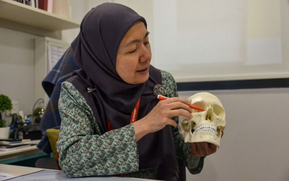
Dr Mah said tumours in the frontal lobe may affect the higher cognitive functions such as personality, social behaviour, impulse control, emotional regulation, movement and speech whereas tumours in the temporal lobe, which processes sound, language, and memory are more likely to result in speech issues and memory loss.
“However, the way these symptoms appear can differ significantly depending on the tumour’s location in the brain. Nearly half of all brain tumour patients experience some form of memory impairment, although the severity often depends on the tumour’s size and exact location,” he said.
To confirm the presence of a brain tumour, Dr Mah said diagnostic imaging tools such as CT scans and contrast-enhanced MRI plays a crucial rule in identifying the tumour’s size, type and exact location within the brain.
“MRI is generally preferred because it provides more detailed images of the brain and can better distinguish between normal and abnormal tissues,” Dr Mah explained. This level of accuracy is vital for planning treatment and determining whether the tumour may be surgically removed.
Dr Gerard Arvind Martin, who is also a Consultant Neurosurgeon at SMCV, noted that secondary brain tumours (those that spread from other parts of the body) occur up to five to ten times more frequently than primary brain tumours, while up to 40 per cent of lung cancer patients may develop secondary brain tumours at some point during their illness.
“Cancers such as lung, breast, colon, and melanoma are most likely to metastasise to the brain, and are commonly seen,” he said, citing a recent and widely reported case in March 2025, in which a local politician revealed that his lung cancer had spread to his brain, resulting in a tumour measuring 3cm.
“The exact reason why certain cancers are more likely to metastasise to the brain remains unclear. This typically occurs when cancer cells break away from the primary tumour, travel through the bloodstream, and lodge in the brain where they begin forming tumoural masses,” he explained.
Dr Gerard Arvind said genetics play a significant role in determining a person’s overall susceptibility to developing brain tumours in their lifetime and in such cases, lifestyle-related factors may act as triggers in individuals who carry these underlying genetic traits, potentially leading to tumour development over time.
Given the uncertainty surrounding brain tumour causes, early detection following symptom development remains the most decisive factor in determining a patient’s outcome.
Once a tumour is diagnosed, its type, size and location will determine the most suitable course of treatment however, surgery remains one of the most effective interventions, particularly for accessible tumours.
A typically recommended procedure is a craniotomy, where a section of the skull is temporarily removed to allow surgeons access to the brain and in selected cases, an awake craniotomy, a highly specialised procedure where the patient is kept responsive during key moments of the operation, may be performed.
“This allows the surgical team to monitor neurological functions such as speech, movement, or vision in real time, helping to preserve critical areas of the brain.” Dr Gerard Arvind explained, adding that for small or hard-to-reach tumours that make open surgery challenging, radiosurgery offers a non-invasive alternative.
Despite its name, it does not involve any surgical incision but uses precisely targeted, high-dose radiation to shrink or control the tumour, often completed in just a single session. Another surgical option in cranial surgery is the use of endoscopes, particularly for tumours located at the skull base or within the pituitary region.
According to Dr Gerard Arvind, the procedure involves inserting a thin, rigid endoscope, equipped with a light and camera, through the nasal passage to access the tumour, which enables surgeons to operate with greater precision and minimal disruption to surrounding tissue.
As a result, patients often benefit from faster recovery times, shorter hospitalisation stay and reduced post-operative discomfort.
“At SMCV, this technique has been successfully employed to treat pituitary tumour cases, often identified by general practitioners or ophthalmologists when patients present with symptoms like visual disturbances and severe headaches,” he said.
Dr Gerard Arvind recounted a successful case in cerebrospinal fluid (CSF) diversion procedures at SMCV involving a lung cancer patient whose secondary brain tumour was obstructing cerebrospinal fluid (CSF) flow, causing life-threatening pressure in the brain.
A minimally invasive procedure called endoscopic third ventriculostomy was conducted to restore the CSF circulation, followed by tumour removal, all in one session and the patient recovered well and was able to proceed with chemoradiotherapy treatment as planned, reflecting how advanced, well-coordinated neurosurgical care can dramatically improve outcomes.
In conjunction with the recent Brain Tumour Awareness Day, it serves as a reminder for the urgent need to prioritise neurological health and to act swiftly when subtle or unexplained symptoms emerge.
Brain tumours may be rare, but their symptoms are real, and often subtle. From constant headaches to mood changes, recognising these signs early can make a critical difference. Early detection not only increases the chances of effective treatment, but also open doors to less invasive procedure and better recover outcomes.
A brain tumour diagnosis is not the end of the road. Thanks to ongoing advancements in diagnostics and minimally invasive surgical techniques, patients today have more options and greater hope than ever before. With timely action and the right guidance and support, recovery is possible, and no one has to face this journey alone.
Was this article helpful?
0 out of 0 found this helpful
Suggest to Read


